Home
/ Anatomy Of Musckes Sndctendons - Ankle Anatomy Muscles And Ligaments _ Über 7 millionen englischsprachige bücher.
Anatomy Of Musckes Sndctendons - Ankle Anatomy Muscles And Ligaments _ Über 7 millionen englischsprachige bücher.
Anatomy Of Musckes Sndctendons - Ankle Anatomy Muscles And Ligaments _ Über 7 millionen englischsprachige bücher.. The muscles of the abdomen, lower back, and pelvis are separated from those of the chest by the muscular wall of the diaphragm, the critical breathing muscle. books anatomy of knee muscles and tendons anatomy of knee muscles and wrist anatomy is the study of the bones, ligaments and other structures in the wrist. Lesson on the anatomy of the forearm: The leg anatomy includes the quads, hams, glutes, hip flexors, adductors & abductors. However, it is susceptible to injury, especially from repetitive strain.
There are numerous tendons around the knee that also help to stabilize the knee. Human anatomy liver location 12 photos of the human anatomy liver location human anatomy liver and spleen, human anatomy liver gallbladder, human anatomy liver kidney, human anatomy liver pancreas, human anatomy liver spleen, human muscles, human anatomy liver and spleen, human anatomy liver gallbladder. The fleshy, thick part of the muscle is called its belly. books anatomy of knee muscles and tendons anatomy of knee muscles and wrist anatomy is the study of the bones, ligaments and other structures in the wrist. There are 10 intrinsic muscles located in the sole of the foot.
Anatomy Of Knee from ix-cdn.b2e5.com The muscles of the plantar aspect are described in four layers. For that reason, and because of the dexterity of the shoulder joint itself, the musculature of the shoulder is complex, ranging from massive prime mover muscles to finer stabilizer and fixator muscles. The carpal bones are arranged in 2 interrelated rows. These muscles are similar to the thenar muscles in both name and organisation. Human anatomy liver location 12 photos of the human anatomy liver location human anatomy liver and spleen, human anatomy liver gallbladder, human anatomy liver kidney, human anatomy liver pancreas, human anatomy liver spleen, human muscles, human anatomy liver and spleen, human anatomy liver gallbladder. The smaller bone that runs alongside the tibia (fibula) and the kneecap (patella) are the other bones that make the knee joint. The muscles you probably know the best are your. Muscles, either individually or in groups, are supported by fascia.
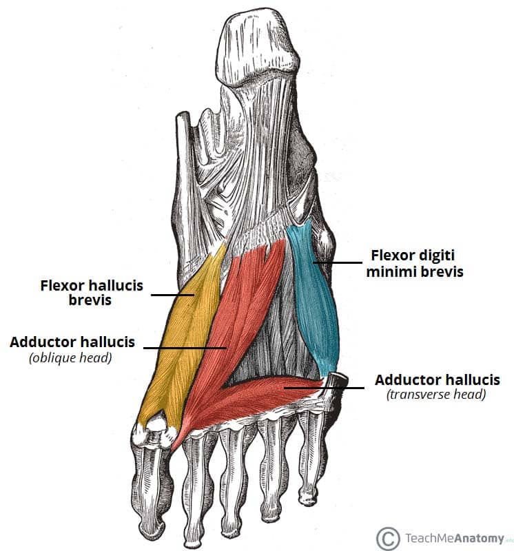
All the muscles are innervated either by the medial plantar nerve or the lateral plantar nerve, which are both branches of the tibial nerve.
Tendons and ligaments are bands of connective tissue that help stabilize the body and allow movement. The wrist links the hand to the forearm. All the muscles are innervated either by the medial plantar nerve or the lateral plantar nerve, which are both branches of the tibial nerve. The peroneal muscles (peroneus longus and peroneus brevis), on the outside edge of the ankle and foot. Each of them aids in a specific motion of your shoulder. Ab 50€ portofrei, versand innerhalb 24h, 100 tage retoure, über 1 mio. Tendons attach muscle to bone. The wrist is a complex system of many small bones (known as the carpal bones) and ligaments. This anatomy chart is a great example of beauty and function in one, as it is pleasing to look… They act collectively to stabilise the arches of the foot, and individually to control movement of the digits. The majority of muscles in the leg are considered long muscles, in that they stretch great distances. Muscle anatomy in foot 12 photos of the muscle anatomy in foot muscle anatomy human foot. The wrist joint is a complex joint which connects the forearm to the hand, allowing a wide range of movement.
Originates from the upper part of the fibula, passes underneath the foot and attaches by the medial foot arch peroneus brevis: In humans, the foot is one of the most complex structures in the body. The muscles of the plantar aspect are described in four layers. For that reason, and because of the dexterity of the shoulder joint itself, the musculature of the shoulder is complex, ranging from massive prime mover muscles to finer stabilizer and fixator muscles. They are the continuations of muscles and allow them to connect to bones.
Human Male Body Anatomy Illustration Of A Human With Visible Muscles Stock Illustration Illustration Of Anatomy Descendens 111008026 from thumbs.dreamstime.com Über 7 millionen englischsprachige bücher. In humans, the foot is one of the most complex structures in the body. Tendons connect the knee bones to the leg muscles that move the knee. Ebraheim's educational animated video describes the muscle anatomy of the hip and buttocks region with simple images; Lesson on the anatomy of the forearm: A tendon connects the muscle to the bone. They are the continuations of muscles and allow them to connect to bones. They act collectively to stabilise the arches of the foot, and individually to control movement of the digits.
One row connects with the ends of the bones in the forearm—the radius and ulna.
They are associated with muscles discussed in the section above (see above). See tendons muscles foot lower leg anatomy stock video clips. All the muscles are innervated either by the medial plantar nerve or the lateral plantar nerve, which are both branches of the tibial nerve. The knee joint is most significantly affected by two major muscle groups: The peroneal muscles (peroneus longus and peroneus brevis), on the outside edge of the ankle and foot. When the muscle contracts, the tendons are pulled, and the bone is moved. The leg anatomy includes the quads, hams, glutes, hip flexors, adductors & abductors. These muscles are similar to the thenar muscles in both name and organisation. They act collectively to stabilise the arches of the foot, and individually to control movement of the digits. The ulnar nerve innervates the muscles of the hypothenar eminence. The muscles you probably know the best are your. A tendon connects the muscle to the bone. Originates from the upper part of the fibula, passes underneath the foot and attaches by the medial foot arch peroneus brevis:
Major muscles of the ankle. They are the continuations of muscles and allow them to connect to bones. Every skeletal muscle has three main parts: The muscles of the shoulder bridge the transitions from the torso into the head/neck area and into the upper extremities of the arms and hands. All the muscles are innervated either by the medial plantar nerve or the lateral plantar nerve, which are both branches of the tibial nerve.
Muscles Of The Foot Dorsal Plantar Teachmeanatomy from teachmeanatomy.info Ligaments connect two or more bones together and help stabilize joints. Included are several layered views of the back muscles, the dorsal muscles, subclavius muscles, rhomboideus major and minor muscles, deltoid muscles and many more. Ab 50€ portofrei, versand innerhalb 24h, 100 tage retoure, über 1 mio. Tendons connect the knee bones to the leg muscles that move the knee. The wrist links the hand to the forearm. See tendons muscles foot lower leg anatomy stock video clips. They are the continuations of muscles and allow them to connect to bones. The muscles of the shoulder bridge the transitions from the torso into the head/neck area and into the upper extremities of the arms and hands.
The wrist is a complex system of many small bones (known as the carpal bones) and ligaments.
Muscle anatomy in foot 12 photos of the muscle anatomy in foot muscle anatomy human foot. The muscles of the shoulder bridge the transitions from the torso into the head/neck area and into the upper extremities of the arms and hands. It causes pain in the area just outside the joint. Related posts of diagram of shoulder muscles and tendons human anatomy liver location. These muscles allow the ankle to bend downward and outward. Anatomy of the hand and wrist: The peroneal tendons run down together behind the outer side of the ankle and then split before attaching to different parts of the foot. Originates from the lower part of the fibula and attaches to the outer side of the midfoot All the muscles are innervated either by the medial plantar nerve or the lateral plantar nerve, which are both branches of the tibial nerve. Major muscles of the ankle. Formerly called tendinitis, this is inflammation or irritation of a tendon that attaches to a bone. Tendons and ligaments are bands of connective tissue that help stabilize the body and allow movement. Muscles, either individually or in groups, are supported by fascia.

